


New Microscope Captures 3D Videos of Components Inside Living Cells
News 2026/11/02
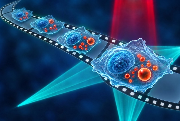
Masato Fukushima (graduate student), Keiichiro Toda (Project Assistant Professor), Yusei Sugawara (graduate student), and Shotaro Kawano (Project Researcher) from the Graduate School of Science at the University of Tokyo, together with Professor Takuro Ideguchi from the Graduate School of Frontier Sciences at the University of Tokyo (also Team Director at the RIKEN Center for Advanced Photonics), and their colleagues have developed a mid-infrared photothermal microscope capable of capturing three-dimensional (3D) videos of chemical components inside living cells. Mid-infrared photothermal microscopy is a label-free imaging technique that visualizes chemical components within cells at high spatial resolution by detecting changes in refractive index (photothermal effects) induced by the absorption of mid-infrared light associated with molecular vibrations, using visible light.
In this study, the researchers accelerated conventional 3D measurements by approximately 400-fold, achieving 3D chemical imaging at a rate of 19.2 volumes per second (equivalent to acquiring 19.2 3D images every second). This capability enabled the 3D tracking of lipid droplets inside living cells based on lipid-specific signals and the quantitative analysis of their motion. The researchers also demonstrated spectroscopic imaging, acquiring 3D images at approximately 20 distinct wavenumbers within one second. These advances establish a new approach for investigating the relationship between intracellular dynamics and chemical composition in three dimensions.
See below for more information.
- UTokyo FOCUS:https://www.s.u-tokyo.ac.jp/en/press/11278/
- Aritcle URL:https://www.nature.com/articles/s41467-026-78699-z
- Ideguchi Lab:https://www.ideguchi.ipst.s.u-tokyo.ac.jp/en/
